
Pancreatic cancer has a five-year survival of less than 11%, a rate which drops even further once the cancer has metastasized. Improvements in treatments are urgently needed. Now, live images of drug response and resistant “pockets” in pancreatic cancer have been revealed by a novel tool developed by researchers at the Garvan Institute. The researchers developed a biosensor mouse model that produces a fluorescent version of the molecule AKT, which was found to cause cancer cells to spread, and provides the first real-time view of a major cancer cell-signaling network within living tissues.
The findings are published in Science Advances in an article titled, “Monitoring AKT activity and targeting in live tissue and disease contexts using a real-time Akt-FRET biosensor mouse.”
“Aberrant AKT activation occurs in a number of cancers, metabolic syndromes, and immune disorders, making it an important target for the treatment of many diseases,” wrote the researchers. “To monitor spatial and temporal AKT activity in a live setting, we generated an Akt-FRET biosensor mouse that allows longitudinal assessment of AKT activity using intravital imaging in conjunction with image stabilization and optical window technology. We demonstrate the sensitivity of the Akt-FRET biosensor mouse using various cancer models and verify its suitability to monitor response to drug targeting in spheroid and organotypic models.”
“The AKT pathway has been a target for therapy for decades but drugs targeting this pathway alone have thus far been largely ineffective in the clinic,” said Paul Timpson, PhD, head of the invasion & metastasis lab, co-lead of the cancer ecosystems program at Garvan, and co-senior author of the study.
The new imaging model will help the researchers see a clear way for how combination approaches could reduce drug resistance and switch off AKT to help stop tumors from growing and spreading.
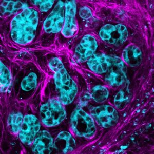
“Using intravital microscopy at the ACRF INCITe Centre to image our biosensor model, we blocked AKT activity using clinical treatments, but could see, in real-time, that AKT remained switched on inside drug-resistant pockets, specifically in areas close to invasive borders or with low oxygen supply,” said co-senior author Max Nobis, PhD, head of the Intravital Imaging Expertise Center at the VIB-KU Leuven Center for Cancer Biology and visiting scientist at Garvan.
“We also saw that as cancer cells got closer to a blood vessel, AKT started to activate. This indicates that AKT not only drives cancer growth, but it also actively drives the early events of cancer spread to other sites around the body. For the first time, we’re able to see these events in a live pancreas.”
The researchers will next use the biosensor model to optimize new combination therapies with AKT pathway inhibitors for pancreatic cancer and other AKT-driven cancers.
“Our ability to see AKT in cancer cells is taking the guesswork out of the development of new therapies and has broad applications, with AKT contributing to many cancers, including breast and prostate cancers. Our model is a vital new tool in the era of co-targeting the tumor ecosystem in precision medicine, bringing us closer to improved treatment options for patients,” said Timpson.
